
Translate this page into:
Theragnosis using fluorescence: A review
*Corresponding author: Neethu C. Annigeri, Department of Periodontology, Bapuji Dental College and Hospital, Davangere, Karnataka, India. neethuannigeri@yahoo.com
-
Received: ,
Accepted: ,
How to cite this article: Annigeri NC, Mohan R, Vijayakumar DB, Lingaraj AJ. Theragnosis using fluorescence: A review. J Adv Dental Pract Res. 2023;2:59-61. doi: 10.25259/JADPR_20_2023
Abstract
Theragnosis is a novel concept in which diagnosis and treatment, along with the monitoring of the therapy given are done. In dentistry, this concept is applied by utilizing the inherent property of the tooth, which exhibits fluorescence. There is a difference in diseased and sound tooth structure along with any bacterial by-product exhibiting fluorescence. Thus, the present review aims to briefly describe the theragnostic approaches in dentistry. With advancements in the diagnostics aids for better therapy and monitoring simultaneously, a revolution can be made in the field of dentistry. Theragnosis, being a novel concept, gives a way for diagnosis, therapy, and monitoring simultaneously allowing for any alteration in the treatment regimen based on patient response. This review would help the clinicians to adopt one of the newer technologies available in the field of dentistry for diagnosis, treatment planning and also monitor the response.
Keywords
Theragnosis
Fluorescence
Diagnosis
Therapy
Monitor

INTRODUCTION
For better diagnosis and treatment, visualization under a proper light source is of utmost importance. For this to be achieved, magnification under such a light source is a significant requirement.[1]
Theragnosis being a novel concept, diagnosis, treatment, and monitoring are done at the same time utilizing a complex system.[2,3] Theragnosis is used in cancer therapy for diagnosis and real-time monitoring of positive or negative responses by the patient for the given theragnostic drug. This allows the clinician to alter the regimen suitably for the individual patient.[4] The emergence of such a concept has made a revolution in the medical field, changing from single use to multifunctional devices.[5]
Theragnostic helps in optimizing drug selection with better molecular understanding. It includes personalized medicine, pharmacogenomics, and molecular imaging. The development of such a technology plays an important role in personalized medicine, wherein medical treatment is tailored based on the characteristics of individual patients.[4,6]
Thus, theragnosis has three main components: Diagnosis, therapy, and monitoring of the response to therapy [Figure 1].[7]
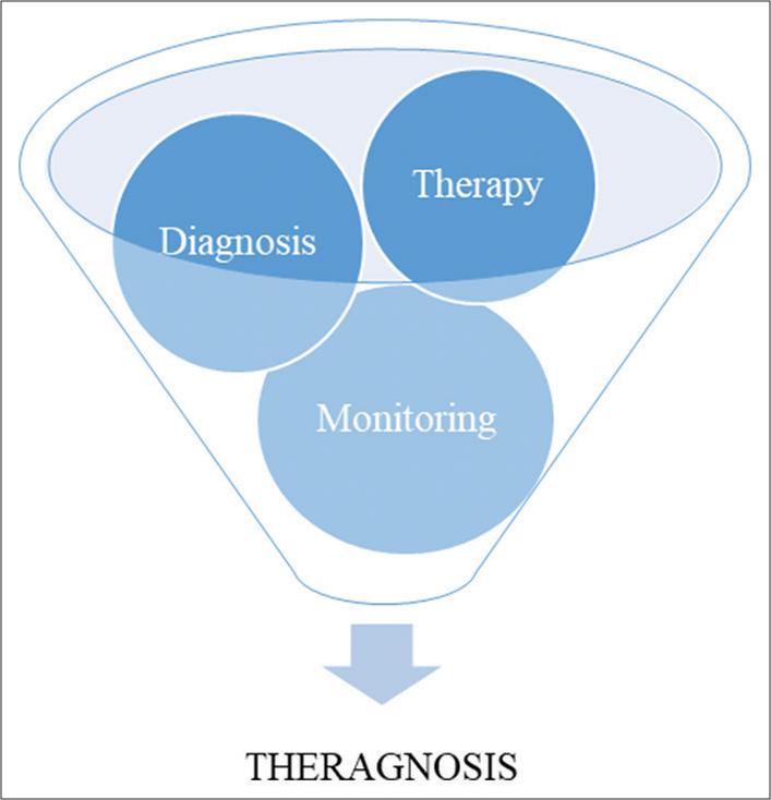
- Theragnosis
THERAGNOSIS IN DENTISTRY
In dentistry, quantitative light-induced fluorescence (QLF) technology based on a 405 nm light source is a multifunctional device used. Such QLF technology is known to be used in the diagnosis of early caries and monitoring remineralization,[8,9] for the detection of cracks in the tooth structure, bacterial deposits such as dental plaque and calculus, for diagnosing peri-implantitis.[1] Recently, such a technology has been used for antimicrobial treatment.[10-12] Furthermore, QLF technology is used for tooth bleaching and evaluation of the efficacy of the tooth bleaching with the use of a 405 nm light source.[3,13-16]
Steier, in 2020, devised a piece of equipment that is wearable and consists of glasses with mounted magnification loupes. For the glasses, the provision for an individual-prescribed eyesight rectification implementation option is provided. The coatings on the glasses and magnification loupes prevent eye injury from emitted light and filter the emitted fluorescence intraorally. Thus, optimal diagnosis and differentiation are achieved.
THE FLUORESCENCE IN THERAGNOSIS
Fluorescence is an emitted radiation of a longer wavelength resulting from the absorption of light of a short wavelength. This effect is due to electrons de-exciting from higher energy levels to lower energy levels. Fluorophores absorb light and get excited electronically to higher energy levels, then de-excite to lower energy levels with the emission of radiation.[17] Fluorescence in dentistry is not a new technology. Stübel, on investigations of fluorescent properties of various body tissues, found the tooth fluorescent blue color second most, with the lens of the eye being first.[18] Using ultraviolet (UV) light, Bommer detected the emission of orange and red colors of dental plaque and calculus.[19] With the same light source, Benedict showed that dentin is more fluorescent than enamel which was due to an organic compound. Furthermore, emission of reddish orange color was seen by supragingival calculus with UV light of longer wavelength.[20]
The emission of fluorescence in all directions is due to fluorophores. This emission intensity is directly proportional to the intensity of the excitation source.[21]
Bacterial byproducts such as porphyrin in the mouth and inherent tooth property (fluorescence) cause photoluminescence when an external source of light in a defined spectrum is used inside the oral cavity. This fluorescence exhibited by enamel is due to modified hydroxyapatite, a carbonate-substituted calcium-deficient apatite. For such visualization, filters are necessary.[1] There is a difference in fluorescence for sound enamel, and after the tooth is caries process initiates and as caries progresses, the fluorescence increases.[22,23] Furthermore, the fluorescence is lesser in whiter teeth as compared to the darker ones.[22]
For caries detection, fluorescence is used since it differentiates between sound and demineralized enamel, which enhances when a light source in the range of 488 nm, that is, blue– green range used.[24-26] The depth of penetration of red light is more, which enables the detection of dentinal caries beneath apparently sound enamel. Carious lesion exhibits more intense fluorescence than the sound one.[27] This is due to more scattering of absorbed light in demineralized tooth structure.[28]
Fluorescence in theragnosis helps in diagnosing the degree of demineralized tooth structure with remineralization potential that requires restorative treatment, and that does not need treatment.[29]
In-office tooth bleaching done with higher concentrations of H2O2 generates free radicals by a complex oxidative process. Reports suggest its damaging action on dental pulp[30] and enamel microhardness.[31] QLF, an additional catalytic source of energy for activation, instead acts by photo-catalytic action for free radical generation by activation with lower concentrations of H2O2.[32,33]
Laser fluorescence is relatively safe compared to any ionizing radiation and has an advantage of accuracy. Thus, more accurately, it can subgingival calculus than with the use of periodontal probing methods.[17] In the case of caries, without an obvious cavity, false-positive and false-negative findings often occur, which could be avoided with the use of fluorescence.[29,34,35]
CONCLUSION
With the evolution in the scientific field, dentistry is evolving in the early diagnosis of various diseases and conditions. Theragnosis is one such evolution which has enhanced the ability for early diagnosis along with therapy and monitoring. The future of theragnosis seems to be more accurate than the current contemporary diagnostic aids available and has the potential in the field of periodontology, restorative, and endodontic dentistry. To know the degree of carious demineralized occlusal surfaces susceptible to remineralizing treatment, the demineralized occlusal surfaces require restorative treatment and the surfaces that do not require treatment. Although in the research arena, advancements are at high velocity, dentists are slow in adopting such advancements in their regular practice for various reasons. Thus, with the utilization of theragnosis in regular dental practice, enhancement in the diagnosis and monitoring of the outcome of given therapy is possible.
Ethical approval
The Institutional Review Board approval is not required.
Declaration of patient consent
Patient’s consent not required as there are no patients in this study.
Conflicts of interest
There are no conflicts of interest.
Use of artificial intelligence (AI)-assisted technology for manuscript preparation
The authors confirm that there was no use of artificial intelligence (AI)-assisted technology for assisting in the writing or editing of the manuscript and no images were manipulated using AI.
Financial support and sponsorship
Nil
References
- Reveal: Fluorescence enhanced theragnosis by designs for vision. Eur J Dent. 2020;14:186-8.
- [CrossRef] [PubMed] [Google Scholar]
- Theranostic nanoplatforms for simultaneous cancer imaging and therapy: Current approaches and future perspectives. Nanoscale. 2012;4:330-42.
- [CrossRef] [PubMed] [Google Scholar]
- Application of quantitative light-induced fluorescence technology for tooth bleaching treatment and its assessment: An in vitro study. Photodiagnosis Photodyn Ther. 2019;25:208-13.
- [CrossRef] [PubMed] [Google Scholar]
- Tumor-targeting multi-functional nanoparticles for theragnosis: New paradigm for cancer therapy. Adv Drug Deliv Rev. 2012;64:1447-58.
- [CrossRef] [PubMed] [Google Scholar]
- Simulation method for developing multiple-use medical devices from re-using and enhancing design of single-use device. Proc CIRP. 2013;5:37-41.
- [CrossRef] [Google Scholar]
- Development of drugs and technology for radiation theragnosis. Nucl Eng Technol. 2016;48:597-607.
- [CrossRef] [Google Scholar]
- Recovery percentage of remineralization according to severity of early caries. Am J Dent. 2013;26:132-6.
- [Google Scholar]
- Development of a fluorescence-image scoring system for assessing noncavitated occlusal caries. Photodiagnosis Photodyn Ther. 2018;21:36-42.
- [CrossRef] [PubMed] [Google Scholar]
- Detection and analysis of enamel cracks by quantitative light-induced fluorescence technology. J Endod. 2016;42:500-4.
- [CrossRef] [PubMed] [Google Scholar]
- Assessing the use of quantitative light-induced fluorescence-digital as a clinical plaque assessment. Photodiagnosis Photodyn Ther. 2016;13:34-9.
- [CrossRef] [PubMed] [Google Scholar]
- Bactericidal effect of the photocatalystic reaction of titanium dioxide using visible wavelengths on Streptococcus mutans biofilm. Photodiagnosis Photodyn Ther. 2017;18:279-83.
- [CrossRef] [PubMed] [Google Scholar]
- The use of QLF to quantify in vitro whitening in a product testing model. Br Dent J. 2001;191:566-9.
- [CrossRef] [PubMed] [Google Scholar]
- The evaluation of a novel method comparing quantitative light-induced fluorescence (QLF) with spectrophotometry to assess staining and bleaching of teeth. Clin Oral Investig. 2010;14:19-25.
- [CrossRef] [PubMed] [Google Scholar]
- A new non-vital tooth bleaching method using titanium dioxide and 3.5% hydrogen peroxide with a 405-nm diode laser or a halogen lamp. Laser Phys Lett. 2008;5:454.
- [CrossRef] [Google Scholar]
- Effect of photochemical activation of hydrogen peroxide bleaching gel with and without TiO2 and different wavelengths. Braz J Oral Sci 2016:393-7.
- [Google Scholar]
- Applications of laser induced fluorescence in dentistry. Int J Dent Clin. 2011;3:38-44.
- [Google Scholar]
- The fluroscence of animal tissues in ultraviolet light. Pflugers Arch Eur J Physiol. 1911;142:1-14.
- [Google Scholar]
- A note on the fluorescence of teeth in ultra-violet rays. Science. 1928;67:442.
- [CrossRef] [Google Scholar]
- Fundamentals and applications of biophotonics in dentistry (biophotonics in dentistry) In: Singapore: World Scientific. 2007.
- [CrossRef] [Google Scholar]
- DIAGNOdent: An optical method for caries detection. J Dent Res. 2004;83(Spec No C):C80-3.
- [CrossRef] [PubMed] [Google Scholar]
- Effect of alteration in organic material of the occlusal caries on DIAGNOdent readings. Braz Oral Res. 2004;18:141-4.
- [CrossRef] [PubMed] [Google Scholar]
- Human teeth with and without dental caries studied by visible luminescent spectroscopy. J Dent Res. 1981;60:120-2.
- [CrossRef] [PubMed] [Google Scholar]
- Early detection of enamel caries by the luminescence excited by visible laser light. Swed Dent J. 1982;6:1-7.
- [Google Scholar]
- Laser-induced fluorescence from sound and carious tooth substance: Spectroscopic studies. Swed Dent J. 1985;9:71-80.
- [Google Scholar]
- Development of a diode laserbased fluorescence caries detector. Caries Res. 1998;32:294.
- [Google Scholar]
- Comparison of laser fluorescence and longitudinal microradiography for quantitative assessment of in vitro enamel caries. Caries Res. 1992;26:241-7.
- [CrossRef] [PubMed] [Google Scholar]
- Comparison between a laser fluorescence device and visual examination in the detection of occlusal caries in children. Saudi Dent J. 2012;24:169-74.
- [CrossRef] [PubMed] [Google Scholar]
- Human pulp responses to in-office tooth bleaching. Oral Surg Oral Med Oral Pathol Oral Radiol Endod. 2010;109:e59-64.
- [CrossRef] [PubMed] [Google Scholar]
- Microhardness change of enamel due to bleaching with in-office bleaching gels of different acidity. Acta Odontol Scand. 2012;70:122-6.
- [CrossRef] [PubMed] [Google Scholar]
- Generation of free radicals and/or active oxygen by light or laser irradiation of hydrogen peroxide or sodium hypochlorite. J Endod. 2003;29:141-3.
- [CrossRef] [PubMed] [Google Scholar]
- Effects of the hydroxyl radical and hydrogen peroxide on tooth bleaching. J Endod. 2004;30:45-50.
- [CrossRef] [PubMed] [Google Scholar]
- A report on the NIH consensus development conference on diagnosis and management of dental caries throughout life. J Dent Res. 2004;83(Spec No C):C15-7.
- [CrossRef] [PubMed] [Google Scholar]
- Comparison between visual examination and a laser fluorescence system for in vivo diagnosis of occlusal caries. Caries Res. 2001;35:421-6.
- [CrossRef] [PubMed] [Google Scholar]


