
Translate this page into:
Unmasking the face: A case report of soft-tissue reconstruction after degloving injury
*Corresponding author: Kumar Saket, Department of Maxillofacial Surgery, Rajiv Gandhi Institute of Health Sciences, Bengaluru, Karnataka, India. saket0410@gmail.com
-
Received: ,
Accepted: ,
How to cite this article: Saket K, Kumari K, Vijapur M, Kattimani V. Unmasking the face: A case report of soft-tissue reconstruction after degloving injury. J Adv Dental Pract Res. 2024;3:19-21. doi: 10.25259/JADPR_14_2024
Abstract
Facial degloving injuries present a complex medical challenge, where the skin and underlying tissues detach from the muscles, fascia, and bones beneath. These injuries pose significant difficulties for surgeons, particularly when complicated by wound infection or tissue necrosis. This case report details the experience of a 26-year-old male patient with a history of trauma, who suffered a facial degloving injury requiring reconstructive soft-tissue surgery. It outlines the challenges encountered during both the surgical and post-operative phases in managing a patient with a full-thickness avulsed flap extending from the lower face to the suprahyoid area of the neck.
Keywords
Avulsion
Degloving
Maxillofacial injuries
Soft tissue
Facial injuries

INTRODUCTION
Degloving soft-tissue injuries are a serious surgical problem, characterized by the detachment of skin and subcutaneous tissue from the muscle and fascia as a result of rapid shearing pressures. They are more frequent in men, who have a greater rate of severe injuries. While these injuries can occur anywhere on the body, the lower limbs, trunk, scalp, and face are most commonly impacted, resulting in variable degrees of skin and soft-tissue loss.
These injuries are classed as either closed/internal or open/external wounds. Delays in treatment frequently result in necrosis of the avulsed skin flap due to a reduced blood supply. Furthermore, the wide variety of types, extents, and severity of degloving injuries makes it difficult to develop standardized care regimens and anticipate results. This article focuses on degloving injuries to the lower face and investigates the variables determining the survival and healing of avulsed soft tissue.[1]
CASE REPORT
A 26-year-old patient presented to the oral and maxillofacial surgery (OMFS) department with an 8-day history of pain and a laceration to the lower lip and chin area of the face, attributing the injury to a road traffic accident while riding without a helmet. Before seeking treatment at Karnataka institute of medical sciences (KIMS) casualty on July 22, 2022, the patient had received primary sutures at a government hospital [Figure 1]. After sustaining the injury, he underwent a tetanus toxoid (TT) injection and was subsequently admitted to the department of OMFS at KIMS. There was no reported history of loss of consciousness, seizures/vomiting, or ear/nasal bleeding. Local examination revealed an oral laceration measuring 4 × 4 cm over the upper lip and 6 × 4 cm over the lower lip and chin region, with noticeable facial asymmetry in the lower face. Eye signs and movements were normal, while palpation indicated stretched skin without step deformity, draining sinus, or fistula. No pus discharge was observed at the site, and intraoral examination revealed a 4 × 4 cm laceration in the lower vestibular region, with negative Gureins and Colomens signs [Figure 2]. Clinical and radiological assessments showed no evidence of facial bony injuries.
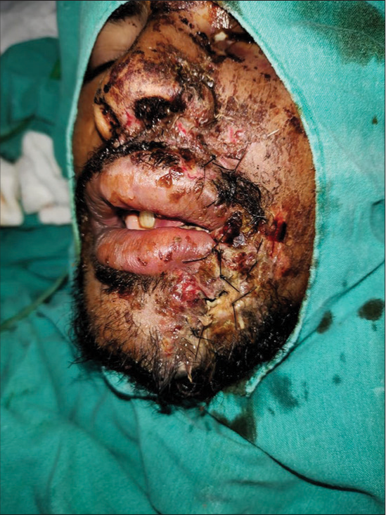
- A 26-year-old male with history of trauma. Pre-operative picture with stay sutures.
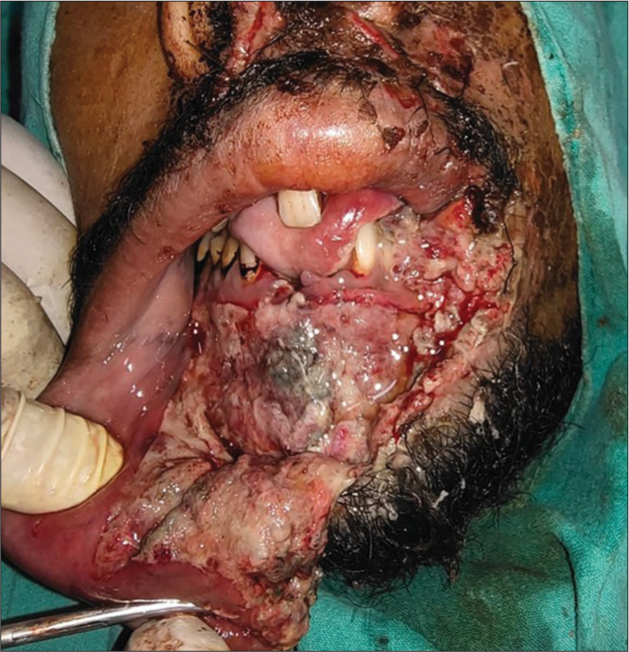
- Image showing degloving injury with necrosed tissues.
Following a comprehensive pre-anesthetic evaluation, the patient underwent general anesthesia through oroendotracheal intubation, with standard face preparation and draping performed. Thorough debridement of the wound was carried out under general anesthesia using normal saline and povidone, excising all devitalized tissues [Figure 3]. The degloved flap was repositioned, and layer-wise suturing with 3–0 vicryl intraorally and 3–0 nylon extraorally was performed to relocate the avulsed structures to their original positions [Figure 4]. Layered suturing of the lower lip was completed, followed by postoperative dressing to prevent hematoma. Due to wound contamination, intravenous injections of cefoperazone + Sulbactam, metronidazole, and paracetamol were administered, along with a tetanus immunoglobulin injection.
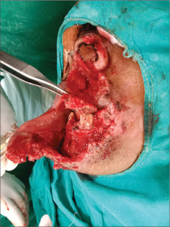
- Image showing debrided and cleaned soft tissues of the lower face.
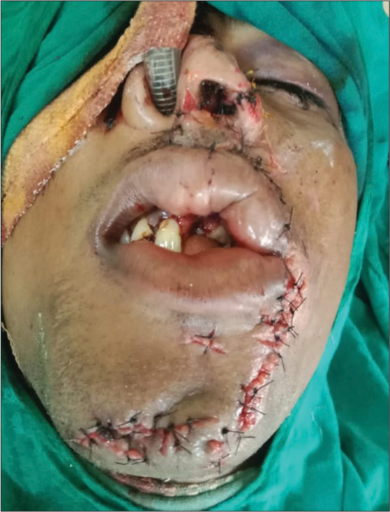
- Immediate post-operative sutured picture.
At the subsequent check-up, the patient reported minor discomfort and tenderness on palpation. Visual inspection revealed anticipated healing of the alveolar mucosa without signs of infection. Sutures were removed, and the patient was advised to schedule follow-up appointments and routine dental check-ups with their regular dentist before discharge. The importance of using appropriate headgear with facial protection and a mouthguard was emphasized to prevent future injuries.
DISCUSSION
The face occupies a singular role in shaping one’s identity and self-worth, surpassing the significance of other bodily features. Consequently, negative repercussions stemming from injury or therapeutic interventions can profoundly impact both the outward appearance and functional aspects of an individual’s life. The central objectives in addressing facial wounds and injuries closely mirror those applied to injuries affecting other anatomical regions.[2] The primary focus lies in the prompt facilitation and consistent maintenance of wound healing processes, coupled with the restoration of alignment and functionality of the implicated soft tissues. This comprehensive approach not only aims to address the immediate physical trauma but also acknowledges the critical role the face plays in interpersonal interactions and self-perception. As such, managing facial injuries entails a nuanced consideration of both esthetic and functional outcomes, striving to optimize the overall well-being and quality of life of the affected individuals.[3]
CONCLUSION
Reconstructing significant facial wounds presents surgeons with numerous challenges. They must navigate the delicate balance between achieving satisfactory aesthetic outcomes and preserving optimal functionality. At present, there are no standardized treatment protocols in place. Delayed intervention for these injuries can increase the risk of infection and necrotizing fasciitis. Moreover, acute injury to the vascular supply may lead to complete flap necrosis.
Ethical approval
The Institutional Review Board approval is not required.
Declaration of patient consent
The authors certify that they have obtained all appropriate patient consent
Conflicts of interest
There are no conflicts of interest.
Use of artificial intelligence (AI)-assisted technology for manuscript preparation
The authors confirm that there was no use of artificial intelligence (AI)-assisted technology for assisting in the writing or editing of the manuscript and no images were manipulated using AI.
Financial support and sponsorship
Nil.
References
- Peterson’s principles of oral and maxillofacial surgery USA: People’s Medical Publishing House; 2012. p. :2000.
- [Google Scholar]
- Fascial space infections In: Bonanthaya K, Panneerselvam E, Manuel S, Kumar VV, Rai A, eds. Oral and maxillofacial surgery for the clinician. Singapore: Springer; 2021.
- [CrossRef] [Google Scholar]
- Infections of the infratemporal fossa: Imaging/clinical correlations. J Otolaryngol. 1991;20(Suppl 3):1-11.
- [Google Scholar]


